A 2018 study reveals novel changes to the brain that may indicate the beginnings of Alzheimer's disease.
Almost anyone over the age of 30 can tell you that their memory is not quite as sharp as it used to be. Whether it’s because the brain is just too crowded with memories from over the years, or the normal decline that goes along with age, most people experience some difference in their memory capability as they tack on the decades.
But what is normal memory loss? For those with a family history of Alzheimer’s, small events of forgetfulness can cause that anxious voice in the back of the head to ask: Is it Alzheimer’s? Forgetting where you put your cellphone or walking into a room only to blank on why you entered in the first place suddenly takes on new meaning. But now, there’s actually a way to see how memory loss takes hold. Scientists have developed a brain scan that can actually see how one type of memory loss might lead to Alzheimer’s.
A study led by the University of California, Irvine, showed that high-resolution functional magnetic resonance imaging (fMRI) can show some of the underlying reasons behind memory loss, revealing how Alzheimer’s interferes with memory recall.
The researchers scanned people’s brains while they asked them to complete two kinds of tasks—an object memory task and a location memory task. The first task asked subjects, comprised of 20 young adults between the ages of 18 and 31, and 20 older adults between the ages of 64 and 89, to identify everyday objects and then compare them to the next object they were shown.
“Some of the images were identical to ones they’d seen before, some were brand-new and others were similar to ones they’d seen earlier—we may have changed the color or the size,” said Michael Yassa, director of UCI’s Center for the Neurobiology of Learning & Memory and the study’s senior author. “We call these tricky items the ‘lures.’ And we found that older adults struggle with them. They’re much more likely than younger adults to think they’ve seen those lures before.”
The next task required subjects to recall where the subject moved from, location-wise—a test older participants fared much better on than the first task.
“This suggests that not all memory changes equally with aging,” said lead author Zachariah Reagh, who participated in the study as a graduate student at UCI and is now a postdoctoral fellow at UC Davis. “Object memory is far more vulnerable than spatial, or location, memory–at least in the early stages.”
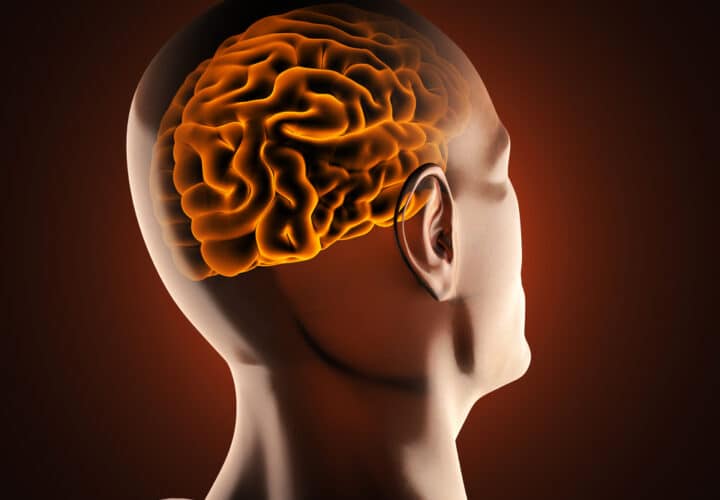
Because they were conducting brain scans as the tests were delivered, scientists were able to pinpoint exactly what happens in the younger brains that doesn’t happen in the older ones. They found that there was a loss of signaling in a place in the brain called the anterolateral entorhinal cortex in older adults. That part of the brain facilitates communication between the hippocampus, the brain’s learning and memory center, and the rest of the neocortex, where the brain stores long-term memories. It’s an area that sees a lot of changes with Alzheimer’s disease, and one of the first that develops beta-amyloid plaques, hardened collections of the toxic protein that spreads across the brain in Alzheimer’s.
“The loss of fMRI signal means there is less blood flow to the region, but we believe the underlying basis for this loss has to do with the fact that the structural integrity of that part of the brain is changing,” said Yassa. “One of the things we know about Alzheimer’s disease is that this region of the brain is one of the very first to exhibit a key hallmark of the disease, deposition of neurofibrillary tangles.”
In the area of the brain that conducts spatial reasoning, there was no age-related difference.
“This suggests that the brain aging process is selective,” Yassa said. “Our findings are not a reflection of general brain aging but rather of specific neural changes that are linked to specific problems in object but not spatial memory.”
Scientists hope that further research will lead to a way to make an early diagnosis for Alzheimer’s patients who are not yet exhibiting symptoms.
“Our results, as well as similar results from other labs, point to a need for carefully designed tasks and paradigms that can reveal different functions in key areas of the brain and different vulnerabilities to the aging process,” said Reagh.
This study was published in the journal Neuron.


