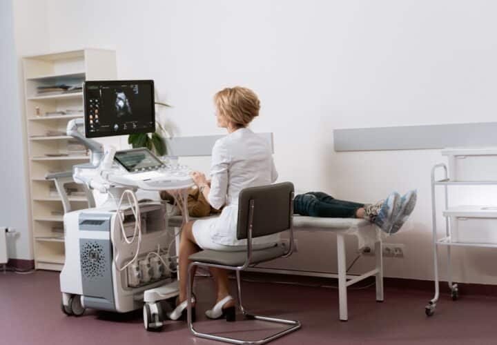
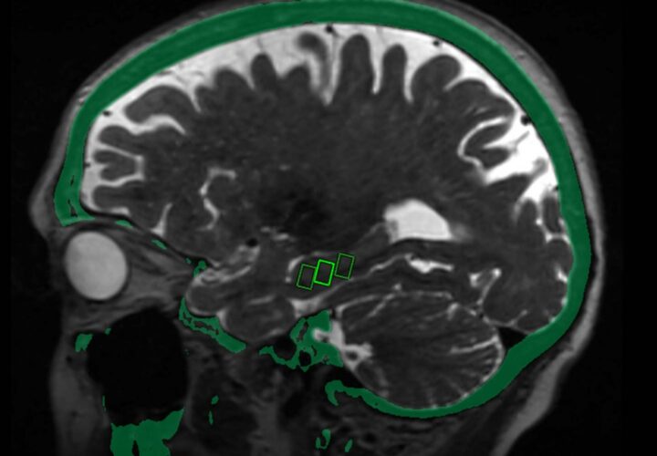
An experimental brain imaging tool explores existing ultrasound technology as a way to detect Alzheimer’s earlier.
Researchers at the University of Illinois, Urbana-Champaign are developing a new imaging tool that could help doctors better study early changes to the brain caused by Alzheimer’s disease. The imaging method — which uses ultrasound — is essentially refining an already existing technique: the use of a microbubble contrasting agent.
The process involves injecting a person with contrast made up of harmless microbubbles, about 10 times smaller than the width of a human hair and filled with an inert gas like perfluorocarbon, that mimic red blood cells and travel through the bloodstream.
Microbubbles work quickly expanding and contracting in response to pressure changes in the sound wave. Ultrasound scanners can then “listen” for these vibrations and translate them into an image. In this case, the image would be of the microvasculature of the brain.
The microbubbles vibrate at such a strong frequency that they create a clearer image of whatever they have been injected to than other contrasts. In other words, they can be used to create clearer images of parts of the brain that have been traditionally hard to see with imaging methods like magnetic resonance imaging (MRI).
How could the imaging help?
According to Dr. Daniel Llano, associate professor of molecular and integrative physiology at the University of Illinois’ Beckman Institute, the new imaging technique could help researchers better learn how things like plaque deposition and microvascular changes are connected to cognitive impairment.
“You can go all the way through the whole brain with very, very high-resolution imaging,” Llano told Being Patient. Llano and Dr. Pengfei Song, an assistant professor in the University’s department of electrical and computer engineering and a faculty member at the Beckman Institute, are spearheading the research behind the new ultrasound imaging technique.
With current imaging techniques, Song says, you can only see “several hundred microns” below the brain’s surface. But with ultrasound imaging, the view goes much much deeper, revealing much more data.
So far, the approach has been tested in mice. But the research, they say, is laying the groundwork to be able to use the technique on humans and perhaps screen for the disease.
While ultrasound imaging allows for better images of structures like the hippocampus, the brain’s memory center, where early signs of Alzheimer’s often appear first, researchers want to focus on using the technique to study blood vessels and their relationship with the disease.
Researchers are particularly interested in using the method to better understand the relationship between plaque depositions, microvascular changes, and cognitive impairment.
This is something that both Song and Llano say is lacking in current Alzheimer’s research.
Next step: more research
Work on the new ultrasound imaging method comes amid calls for more affordable and accessible Alzheimer’s diagnostic tools. But while the possibilities with the method are exciting, Llano and Song don’t believe it will be able to provide all the answers researchers and healthcare providers need to treat Alzheimer’s.
Instead, once the technique has been tested and FDA approved for use on people, it will be an additional diagnostic and research tool for doctors.
“Alzheimer’s is a very complex disease,” said Song. “I don’t think one imaging modality can tell us all the answers to all the questions… The thing we are really working on is filling a hole in the landscape of all the existing biomedical imaging applications.”
Llano and Song’s team recently received a $421,000 grant from the National Institutes of Health to fund two years worth of exploratory and early-stage research.


My wife is late stage alz; I’m heartbroken. But as a retired scientist, I realize that path breaking research will eventually lead to earlier understanding and eventually to a cure. Such a wonderful outcome will lead to longer and healthier lives.