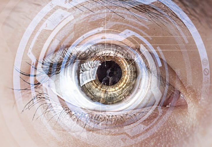
Scientists found that deposits of fat and calcium under the retina could be indicators of Alzheimer’s.
Is the health of the brain reflected in the eyes? New research shows that small deposits in the eyes, formerly thought to be harmless, could actually be an indication of dementia.
Researchers at Queens University in Belfast, Northern Ireland, scanned the eyes of 117 patients between the ages of 60 and 92. They were looking for those yellow spots, known as hard drusen, which are deposits of fat and calcium that form underneath the retina’s top layer. They scanned their eyes once at the beginning of the study and again after two years. An increase in the number of drusen that formed was associated with an increased risk of Alzheimer’s disease. They also discovered that 25 percent of people with Alzheimer’s have hard drusen, compared to 4 percent of people without Alzheimer’s.
“In the peripheral retina, hard drusen accumulation was significantly associated with positive Alzheimer’s disease status,” said lead study author, Dr. Imre Lengyel, in a statement.
More than one expert has suggested that the eyes are the window into brain health. Peter Snyder, Ph.D., Senior Vice President and Chief Research Officer at Lifespan Hospital System, told us that one day, an eye exam could be all doctors need in order to diagnose Alzheimer’s. “The retina is really an extension of our central nervous system,” Snyder said. “It’s often referred to as a protrusion of the brain.”
A previous study showed that beta-amyloid, the toxic protein that accumulates into plaques in the brains of Alzheimer’s patients, was measurable in the eye the same way that it is measurable in the brain.
The researchers who worked on the drusen study hope that this information will lead to a simple eye scan to detect Alzheimer’s, which could lead to a much faster diagnosis at a cheaper and less invasive cost to the patient. Right now, the only way to diagnose a living patient with Alzheimer’s is with a PET scan or taking cerebrospinal fluid, both of which cost thousands of dollars—so much, in fact, that most patients never get them, instead relying on symptoms to diagnose the disease.
The study also found that Alzheimer’s patients have significantly wider blood vessels in the eyes than those without Alzheimer’s. They theorized that the wider vessel made blood flow slower, which could be reflective of what is happening in the brain.
“Our gut feeling is that the eye is mirroring what’s occurring in the brain. Although it’s not exactly the same, it’s the same type of change,” Lengyel told The Daily Mail.
Blood vessel damage in the brain is a hallmark of Alzheimer’s disease.
This study was published in the journal Opthalmic Research.


