Windows to the Brain: Retinal Testing for Alzheimer’s

What if detecting Alzheimer's 20 years ahead of symptoms was as simple as a quick eye test? Dr. Peter Synder shares insights.
Research into Alzheimer’s detection has looked into a variety of possibilities, from blood tests to expensive and extensive brain scanning. But some researchers have found promise in an unlikely location: retinal imaging through the eye. After all, Dr. Peter Snyder tells Being Patient, retinal tissue is an extension of brain tissue, and it can provide clues about brain abnormalities. Plus, retinal scans for Alzheimer’s may be a much more affordable, accessible, and faster way to diagnose the disease — not to mention a less invasive one.
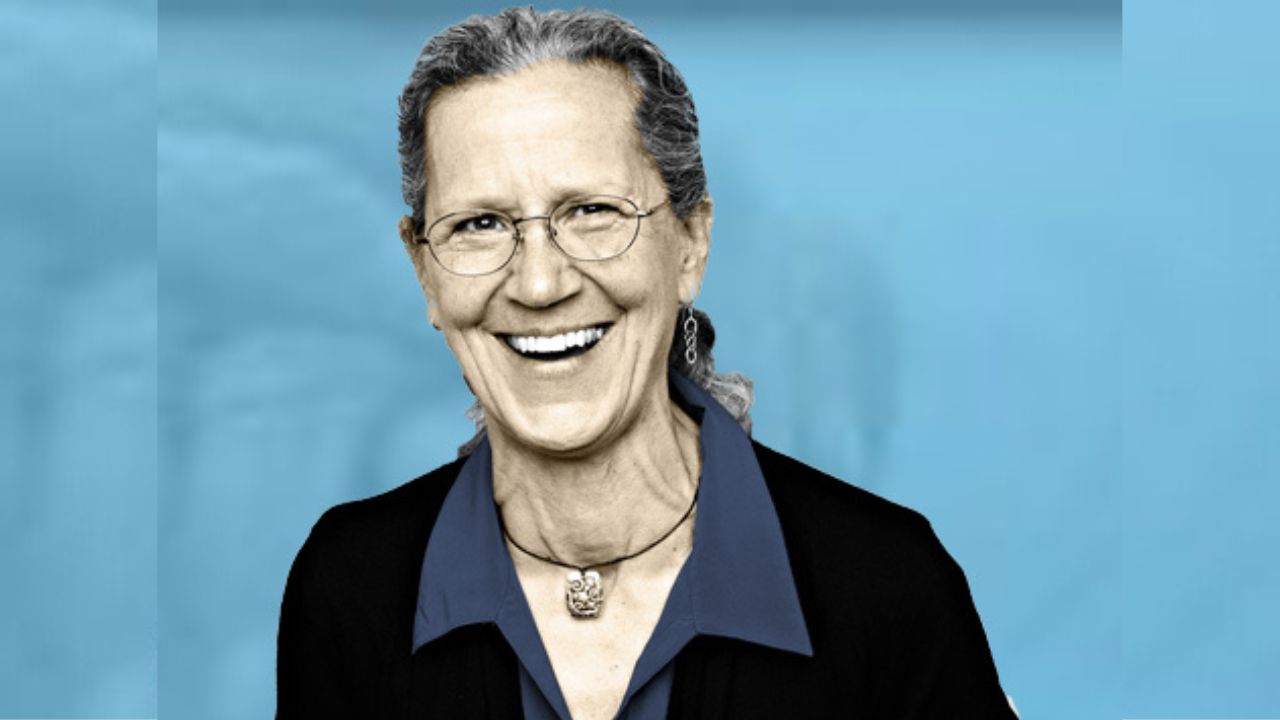
Being Patient spoke with Snyder, a professor of biomedical sciences at the University of Rhode Island, about his research into retinal scanning technology that could detect signs of Alzheimer’s as early as 20 years before symptoms appear.
Why is the eye useful for early Alzheimer’s detection?
Being Patient: When we’re talking about neurodegenerative diseases, what exactly are you looking for through the eyes?
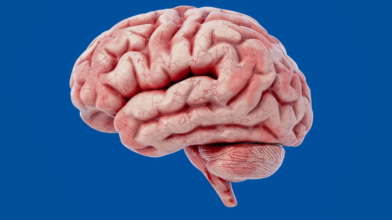
Dr. Peter Snyder: Well, just to step back for a moment, what we’re really interested in is not the entire structure of the eye, but just the retina, because the retina is an extension of the brain. It’s literally a part of our nervous system: It contains the same neurons, the same glial cells, the same vascular supply, the same chemistry as our brains do.
And in fact the cells that form the retina migrate off of the same cluster of cells that form the brain early in embryologic development. So we’re looking at the same tissue. But the really exciting thing about looking at the retina for clues about whether someone may have a brain disease like Alzheimer’s is because it’s exposed tissue and we can look at it in microscopic detail without causing any harm or discomfort, and without being invasive.
So it’s a rare opportunity to look at actual nervous system tissue for clues to the disease—at a potential cost that’s much, much lower than brain scans—as a part of peoples’ daily lives. As people get older, they come into more regular contact with ophthalmologists, and my hope is that these could be points of contact where we can screen many people at a population level in a way that’s affordable.
Being Patient: What is it about the eye that makes it a good spot for early detection?
Dr. Peter Snyder: Some of these systems that seem to be going awry early in the brain, such as changes in the cholinergic system—which is a chemical system that signals between neurons and that we know is affected very early in the disease—we know where that system is in the retina, and we can see it.
We know what layer of cells that chemistry is most active in, and in fact we have seen early changes in that layer in a past study, which led to us starting this new trial. We found in that layer, in the pre-clinical stages of the disease before the onset of symptoms, evidence of increase volume in the structure, which we think represents an inflammatory process.
And we know that inflammation in the brain is a key component in the development of Alzheimer’s disease, so we’re finding some evidence of that in the retina. We also found what we’re calling occlusion bodies aggregating right in that same layer, which we think are blotches of tissue that contain beta-amyloid protein, that are interfering with the transmission of light from the scanner that we’re using.
So we need to follow up on that as well. It may be that there are several things that we’re looking at.
The potential of retinal technology in diagnosing Alzheimer’s
Being Patient: Would it be possible to see through a scan something like beta amyloid and plaque?
Dr. Peter Snyder: Yes, we believe so, but we’re not sure that that’s necessarily going to be the best metric, and it may depend on the stage of the disease. The technology is advancing very rapidly, and we can get beautiful images, but we have to play catchup now to figure what do they mean.
These are images that we have to interpret reliably, and we have to understand what the best quantitative metrics are that we can pull from those images so that when we see a signal we can rely on it, it makes sense, it’s repeatable, and it’s sensitive enough.
Being Patient: What do you think specifically we might be able to get out of these types of tests?
Dr. Peter Snyder: I think we’re developing a screening test to know when older adults in our community need to be referred to a specialty center for more diagnostic, confirmatory tests. A screening test is not the same as making a confirmatory diagnosis; we allow a little bit of false/positive error in these tests because we’d rather catch more people who we’re worried about than not find individuals who may go on to have the disease.
So screening tests may identify persons at risk who the ophthalmologist may say to one day, “I’ve been following you for several years, I’m seeing some changes in your retina that indicate to me a need to refer you to neurologist or a memory disorder center in order to get another round of testing.” That would be a success, because right now we don’t have anything that does that.
I believe that we’re not going to be able to slow progression of the disease, we’re not going to be able to develop a therapy that really is effective, unless we intervene earlier than we’ve attempted so far.
Being Patient: One of the fears in the community about early detection tests is that if someone were diagnosed in their thirties and knew they’d develop Alzheimer’s, that might ruin their life.
Dr. Peter Snyder: Sure, but we do want to pick it up early. I’d like to pick it up a decade or two decades early. We’ve spent decades trying to develop therapies that reverse the damage that’s already been done.
But once someone is clinically showing symptoms, this disease has already been active for up to decades. And I think we’re asking too much of therapeutics to try to undo damage, rather than preventing it. These drugs can only do so much; we can’t reverse time on the damage caused to our brains.
I do think we need to intervene early and identify risks, and you know you’re right, it’s a chicken and egg problem, but ultimately my goal is to protect quality of life. It’s so much easier to protect it than to try and go back.
On the right path
Being Patient: How long before you have conclusive evidence that this type of early detection works?
Dr. Peter Snyder: From our last study that we finished, which was the five-year trial, we have some very exciting evidence that we’re on the right path, we really believe this. So this next trial is to confirm it, and to narrow down the range of metrics to identify the one, two, or three ways of scoring the retinal imaging that we can then translate into a test that we could roll out broadly to optometry and ophthalmology practices.
That’s going to take about a five-year study, and we’re enrolling about a hundred people who we’re going to follow for three years each. And I think three or four years from now we’ll be hot on the path to finding the metric or two that is most important and will develop into a useful tool.